SKU: 50122New
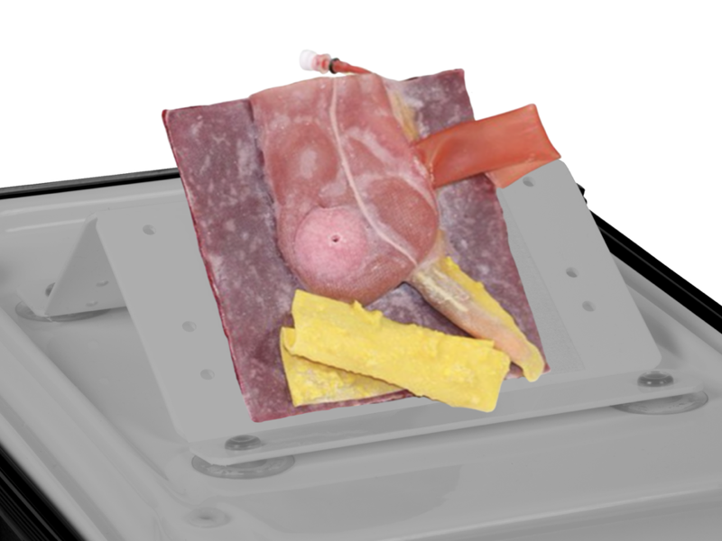
Vermiform Appendix - Normal Anatomy
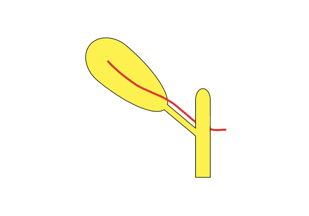
One of a series of three models representing the different locations in which the vermiform appendix is to be found. Trainees will move on from the Surgical Dissection Pads to this progressive 3 stage appendices suite: Normal represents 32% of locations Post-ileal represents 1% of locations Retrocecal represents 64% of locations
One of a series of three models representing the different locations in which the vermiform appendix is to be found. Trainees will move on from the Surgical Dissection Pads to this progressive 3 stage appendices suite: Normal represents 32% of locations Post-ileal represents 1% of locations Retrocecal represents 64% of locations
- The level of difficulty can be increased by progressing from this model on to the Post-ileal and Retrocecal models (not included)
- Convincing fluid filled vessels, cecum contents and tissues encourage correct handling of tissue
- Vessel incorporates a luer lock to which the Mock Blood Giving Set may be attached to provide fluid flow for the appendicular artery. Use Mock Blood - Arterial (250ml)
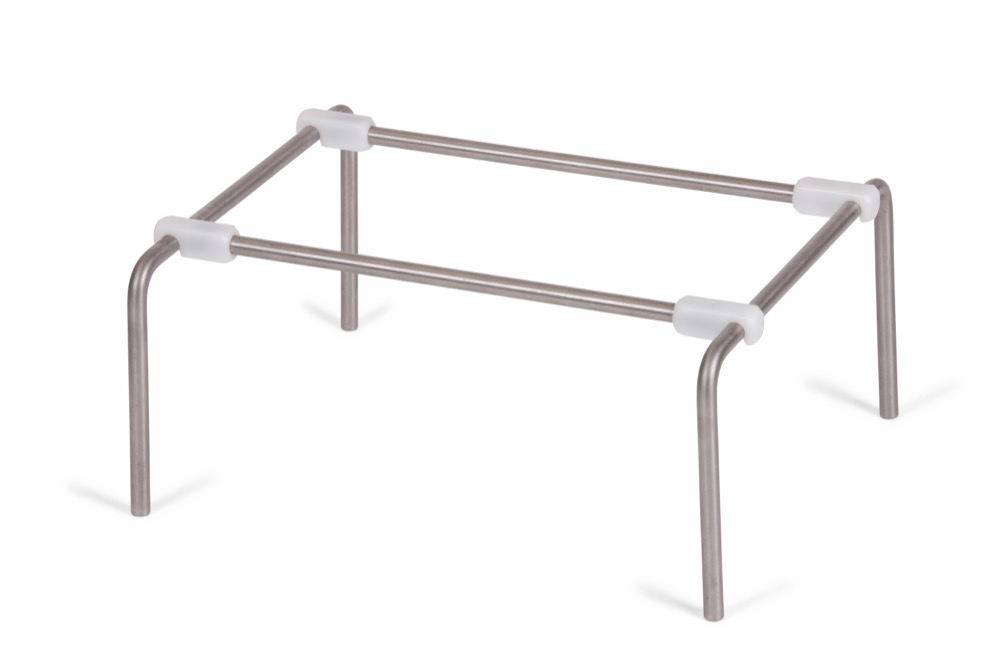
- Ideal for training course requirements; the model is designed for swift attachment to and removal from the Soft Tissue Retaining Set
- Contains latex
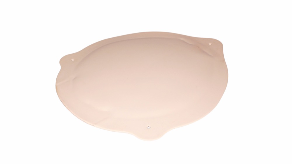
- Highly realistic model of appendix, cecum, and ileum stump presented anatomically
- Excision and division of peritoneum
- Identification and exposure of appendix
- Mobilization and division of vessels
- Removal of appendix
- Inspection of stump of appendix
- Repair and closure of peptic ulcer
No documentation added yet.
Related products